Barrett’s Esophagus
What is Barrett’s esophagus and how is it treated?
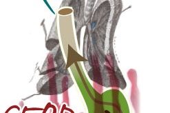
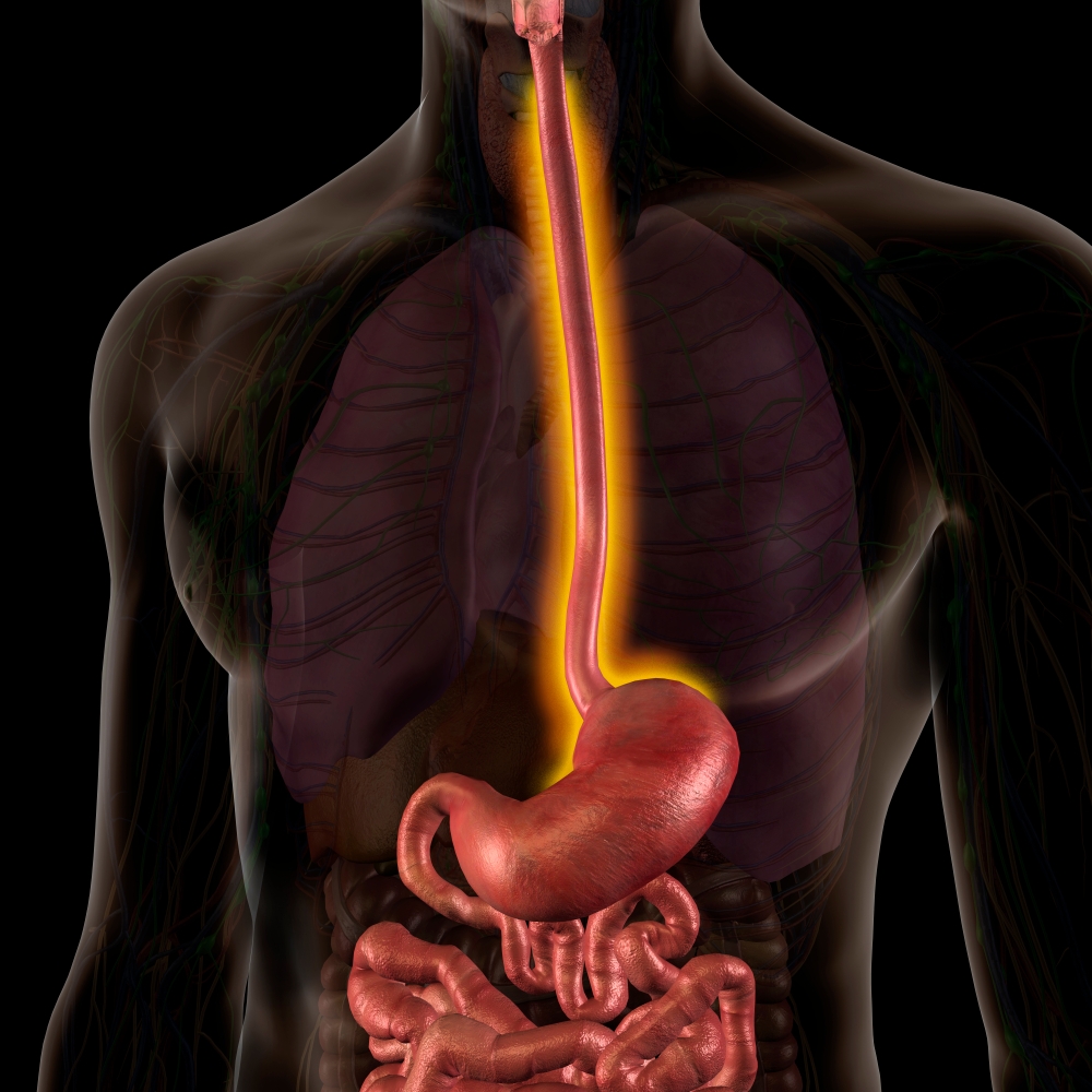
Barrett’s esophagus is a pathological change in the lining of the lower esophagus. It is believed to develop primarily as a result of chronic exposure of the esophagus to gastric acid reflux.
Chronic exposure of the esophagus to stomach acid and other unknown factors results in recurrent mucosal injury. This injury is accompanied by inflammation and, eventually, a cellular change (metaplasia) to a specialized type of columnar epithelium.
Some patients with Barrett’s esophagus may also develop additional precancerous changes called dysplasia. Over time, a small percentage of patients with Barrett’s esophagus may develop esophageal cancer. Your doctor may recommend radiofrequency ablation to treat Barrett’s esophagus with dysplasia (low-grade or high-grade) to reduce the risk of esophageal cancer.
Barrett’s esophagus refers to changes in the macroscopic and microscopic appearance of the inner wall of the esophagus in the area where it connects to the stomach. It is caused by chronic reflux of stomach and small intestine contents into the esophagus.
Barrett’s esophagus is a precancerous condition that can lead to the development of esophageal cancer in some patients. The risk of developing cancer is approximately 0.12 to 0.27% annually. There are several risk factors for the development of Barrett’s esophagus, including:
- Symptoms of gastroesophageal reflux disease (GERD)
- Obesity
- Age (increases with age)
- Gender (male)
- Family history
Patients with Barrett’s esophagus may have no symptoms other than those associated with GERD. These include a burning sensation below the chest and acid regurgitation. Some people with GERD may also have difficulty swallowing.
This symptom requires immediate medical evaluation. These symptoms generally improve with medications that reduce stomach acid.
The only way to confirm the diagnosis of Barrett’s esophagus is through a test called upper endoscopy (gastroscopy). This involves inserting a small, thin, lighted tube (endoscope) through the throat into the esophagus to look for changes in the inner lining (mucosa) of the esophagus.
While the appearance of the esophagus may resemble Barrett’s esophagus, the diagnosis must be confirmed with small tissue samples (biopsy) taken through the endoscope. The pathologist will examine the tissue to make the diagnosis.
Treatment of Barrett’s esophagus is similar to the treatment of GERD. This includes lifestyle changes, such as avoiding certain foods, not eating late at night, and quitting smoking, combined with the use of medications that reduce acid production in the stomach.
Patients with Barrett’s esophagus need medications to reduce acid, such as omeprazole, lansoprazole, pantoprazole, rabeprazole, and esomeprazole. These medications are usually taken before breakfast once a day or, in some cases, before breakfast and dinner. Medications known as H2 receptor antagonists are not as effective in reducing acid in the esophagus caused by Barrett’s esophagus, but they may relieve symptoms for some patients.
Radiofrequency ablation is offered for patients with dysplasia. Radiofrequency ablation (radio waves) is performed through a catheter in the esophagus to destroy the abnormal tissue while minimizing damage to the healthy tissue of the esophagus.
While the patient is under sedation, a device is inserted through the mouth into the esophagus to deliver a controlled level of energy and power to remove a thin layer of diseased tissue. The ability to provide a controlled amount of heat to the diseased tissue is a mechanism by which this treatment has a lower complication rate compared to other forms of therapeutic intervention.
Larger areas of pathological Barrett’s tissue are treated with a balloon catheter. Smaller areas are treated with a tapered end of the catheter. Both catheters are inserted through the endoscope during the gastroscopy, where the thin, flexible tube is inserted through the patient’s mouth.

 What is esophagitis?